近期,德国亥姆霍兹中心金属生物材料研究所研究员Regine Willumeit-Römer在科爱创办的期刊Bioactive Materials发表文章。肿瘤细胞转移是导致癌症病人死亡的主要原因之一。近期,有研究表明含Mg的材料具有抗肿瘤活性。本研究主要探索了含镁材料在肿瘤转移不同阶段中的作用,包括肿瘤细胞迁移、侵袭及肿瘤相关血管生成。实验证明Mg和Mg-6Ag材料均可有效降低骨肉瘤细胞迁移和侵袭能力,并在乏氧环境中减少肿瘤相关血管的生成。
引言
“癌症转移”是指原发灶的肿瘤细胞在远处形成新的继发性肿瘤,极大降低了癌症患者的治愈率和生存率,因癌细胞转移而死亡的比率占癌症死亡率的90%以上。化疗和放疗通常用于治疗肿瘤原发部位残留癌细胞的扩散,但仅对少数患者有效,且副作用明显。因此,需要新的局部治疗策略抑制肿瘤细胞扩散,以提高癌症治疗的成功率和患者生命质量。近期,研究表明一种可降解的含镁的材料表现出明显的肿瘤细胞毒性,且具有良好的生物相容性,因此,可作为载药体系递送抗肿瘤活性药物。该材料的降解速率可改变肿瘤细胞微环境,如pH值、渗透压、氢气含量等,从而抑制肿瘤细胞生长。当含镁材料中引入银(Ag)或其他金属元素会改变材料的降解率和生物效应,如加入Ag可增加该材料的抗菌活性。此前,研究报道了补充Mg2+在肿瘤发生发展中的双面性,既能促进早期肿瘤的生长,也能抑制晚期实体瘤的侵袭和转移。然而,含镁材料是否具有相同的抗肿瘤活性尚未被研究。本研究着重探索了含镁材料对肿瘤相关血管生成、肿瘤侵袭和转移的作用。
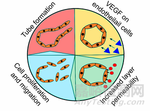
在临床上,骨肉瘤诊断表明有15-20%的转移率,伴随着高死亡率和严重并发症。肿瘤细胞的转移过程伴随着上皮间充质转化(EMT),E-钙粘素(E-cadherin)蛋白下调,及波形蛋白(vimentin)和基质金属蛋白酶(matrix metalloproteinases, MMP)的上调。在肿瘤微环境中,癌细胞与癌旁组织细胞发生密切的信号交流以诱导肿瘤细胞的持续转移,例如成纤维细胞和巨噬细胞。肿瘤细胞经常利用癌旁组织释放的金属蛋白酶MMP-2和MMP-9来降解癌细胞周围的基质,促进肿瘤细胞进入到血管中,随血液循环到达新的部位产生继发性肿瘤。恶性肿瘤快速的增殖需要血管运输大量的氧气和营养物质,当肿瘤中心区域缺氧(O2<3%)且营养不良时,就会导致肿瘤血管异质性。研究表明,血管内皮生长因子(vascular endothelial growth factor, VEGF)是血管生成最重要的因子之一,几乎参与了血管生成的所有步骤。排列在肿瘤血管壁上的内皮细胞之间间隙增大,有利于肿瘤细胞渗透到血管内,同时内皮细胞也会增殖和迁移以形成性的肿瘤血管,并与新的内皮细胞发生连接(如图1所示)。本研究内容表明,Mg2+可显著影响骨肉瘤细胞的迁移和侵袭,并改善肿瘤血管异质性。
图1 常规、聚乙二醇化、靶向和多功能脂质体的简化表示
1. 镁基材料减少了癌细胞的迁移和侵袭
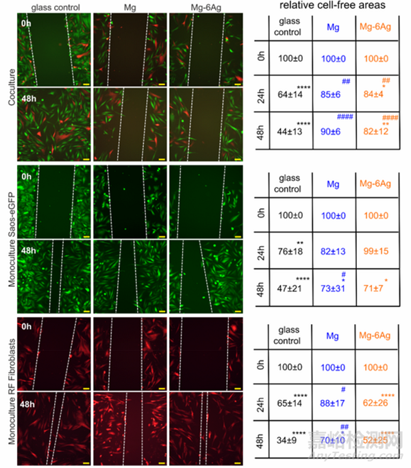
将骨肉瘤细胞(Saos-eGFP)和成纤维细胞单独或共同接种于细胞培养皿或覆盖了Mg或Mg-Ag材料的培养皿中培养0-72 h。通过划痕实验观察不同细胞在不同培养环境中的迁移能力。如图2所示,当成纤维细胞与骨肉瘤细胞共同存在时,Mg或Mg-Ag材料可显著抑制细胞的迁移。当单独培养细胞时,Mg或Mg-Ag材料也能明显抑制癌细胞的迁移,但抑制成纤维细胞迁移的能力下降。
Fig. 2. Cell migration influenced by Mg and Mg–6Ag under normoxia. Microscopic images of Saos-eGFP (green) and RF Fibroblasts (red) in coculture or monocultures with the initial wound (0 h) and after 48 h. The white dotted lines symbolize the cell fronts. Scale bar is 100 μm. The cell-free areas after 24 h and 48 h werequantified in relation to the initial cell-free area. Relative cell-free areas are shown as the mean ± SD from two experiments with two samples and three randomlychosen positions (n = 12). Statistics: two-way ANOVA (Mg, Mg–6Ag compared to glass control = #; 24 h, 48 h compared to 0 h = *) with Tukey’s multiplecomparison test. One symbol = p < 0.05; two symbols = p < 0.01; three symbols p < 0.001; four symbols = p < 0.0001.
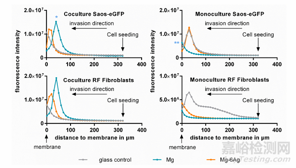
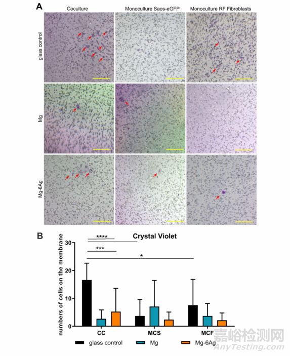
此外,为研究含镁材料对癌细胞侵袭能力的影响,作者将骨肉瘤细胞(Saos-eGFP)和成纤维细胞单独或共同接种于带有ECM凝胶的小室中,底部加入细胞培养基或经过Mg或Mg-Ag材料处理的培养基,通过检测细胞向下侵袭的荧光强度来代表细胞的侵袭能力。图3表明,骨肉瘤细胞和成纤维细胞共同接种于含Mg材料或Mg-Ag材料的转移小室中,两种细胞的侵袭能力均明显降低;但两种细胞单独与含Mg材料或Mg-Ag材料共培养时,细胞的侵袭能力与对照组相比稍有增加。图4对图三中穿过ECM的细胞进行结晶紫染色,并得出一致结论。
Fig. 3. Cell invasion influenced by Mg and Mg–6Ag. Cells invaded through an ECM mimetic gel layer from the point of cell seeding (320 μm) to the membrane (0μm). Fluorescence intensities of representative z-stack (10 μm steps) images are shown. Statistics: ordinary one-way ANOVA with Dunnett’s multiple comparison test(n = 12); * = p < 0.01, ** = p < 0.01.
Fig. 4. Visualization of invasive cells. (A) Cells that invaded the ECM mimetic gel layer and crossed the membrane were stained with crystal violet. Red arrows onrepresentative images indicate invaded cells, which were quantified (red numbers). Scale bar is 100 μm. (B) Quantitative analysis of images. Statistics: two-wayANOVA (Mg, Mg–6Ag compared to glass control = *) with Tukey’s multiple comparison test (n = 12). * = p < 0.05; *** = p < 0.001; **** = p < 0.0001.
综上,骨肉瘤细胞和成纤维细胞与含镁材料共孵育时,骨肉瘤细胞的迁移和侵袭能力均被明显抑制,但骨肉瘤细胞单独与含镁材料共培养后,癌细胞的迁移和侵袭能力并未下降甚至稍有增加。
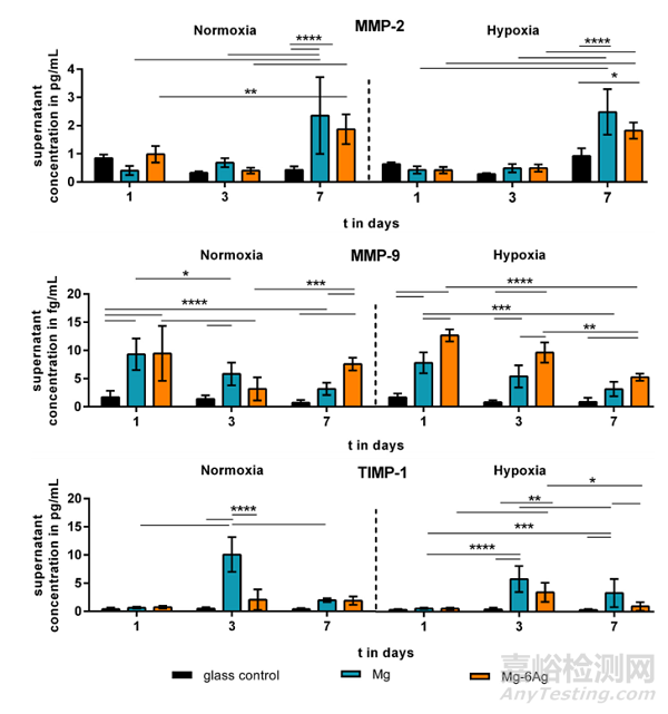
2. 含镁材料能促进MMP-2、MMP-9和TIMP-1的释放
肿瘤组织中高水平的金属蛋白酶会导致ECM降解,促进单个肿瘤细胞迁移并侵袭进入到血管中。图5显示Mg或Mg-Ag材料共培养肿瘤细胞会促进MMP-2、MMP-和TIMP-1分泌增加,共培养7天后,MMP-2的分泌量是对照组的2-3倍;MMP-9在含镁材料的共培养条件下也显著增加,但随共培养的时间呈下降趋势;而TIMP-1仅在第三天有明显增加,且Mg共培养的效果优于Mg-Ag。
Fig. 5. The impact of Mg-based materials onmetastases-associated cytokine release. MMP-2,MMP-9 and TIMP-1 was quantified in the supernatant of migrating cells of the coculture and normalized to the cell numbers. Normalized cytokineconcentrations are shown as the mean ± SD from twoexperiments with two samples in duplicates (n = 12).Statistics: two-way ANOVA (materials, time points)with Tukey’s multiple comparison test. * = p < 0.05;** = p < 0.01; *** = p < 0.001; **** = p < 0.0001.
3.含镁材料减少肿瘤血管生成
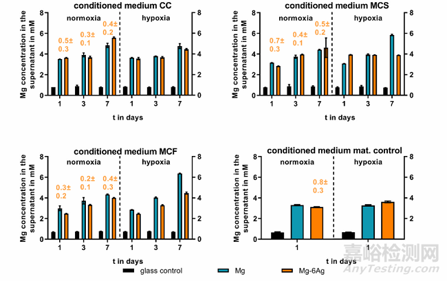
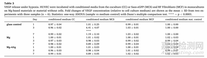
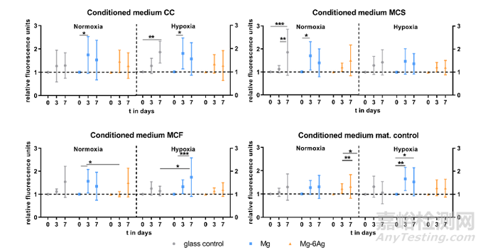
用HUVEC间接模型研究癌症诱导血管生成。首先,如图6所示,普通培养基中Mg2+的浓度约为0.8 mM,实验组培养基分别加入含可降解的Mg和Mg-Ag材料。随与细胞共孵育时间的增加,培养基中游离的Mg2+浓度逐渐升高到3-6 mM,且Mg2+的释放与氧气浓度和孵育细胞无关。研究表明,肿瘤相关血管生成是由ECM的降解内皮细胞的上皮间质转化导致的。图7探索了含镁材料和不含镁材料条件对人脐静脉内皮细胞(HUVEC)的影响。常氧条件下,第3-7天内皮细胞间连接通透性明显增加,而在缺氧条件下改变不明显。在第7天,HUVEC与来自对照组的细胞的条件培养基(CC、MCS、MCF)孵育时,其渗透性略高于与含镁材料的条件培养基共培养组。VEGF是血管生成最重要的介质之一,可形成新血管。因此,测量了条件培养基培养HUVEC后的上清液中VEGF浓度(Table 3)。含镁材料培养基和对照培养基共培养并未引起明显不同的VEGF释放,相反,HUVEC与低氧条件下对照组的培养基孵育,可在第3天显著增加VEGF浓度。
Fig. 6. Supernatant Mg and Ag concentration of conditioned media. Saos-eGFP and RF Fibroblasts were seeded as a 1:1 coculture (CC) or monocultures (Saos-eGFP:MCS, RF Fibroblasts: MCF) on Mg, Mg–6Ag or glass (glass control). Furthermore, material without cells served as a mat. control. After one, three and seven days,conditioned medium was harvested, and Mg (in mM) and Ag (orange numbers, in μM) concentration were measured as described previously.
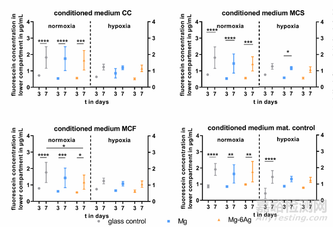
Fig. 7. Endothelial cell layer permeability withdifferent conditioned media. HUVEC were incubatedwith conditioned media from the coculture (CC) orSaos-eGFP (MCS) and RF Fibroblasts (MCF) inmonocultures on Mg-based materials or materialwithout cells. Fluorescein-dextran concentrations (inμg/mL; quantified with a standard curve) are shownas the mean ± SD from two experiments with onetriplicate (n = 6). Statistics: two-way ANOVA (materials, time points) with Tukey’s multiple comparisontest. * = p < 0.05; ** = p < 0.01; *** = p < 0.001;**** = p < 0.0001.
Table 3VEGF release under hypoxia. HUVEC were incubated with conditioned media from the coculture (CC) or Saos-eGFP (MCS) and RF Fibroblasts (MCF) in monocultureson Mg-based materials or material without cells. Fold changes of VEGF concentration (relative to cell culture medium) are shown as the mean ± SD from two experiments with three samples (n = 6). Statistics: one-way ANOVA (sample vs medium control) with Dunn’s multiple comparison test. **** = p < 0.0001.
4. 含Mg材料影响内皮细胞增殖、迁移和血管再生
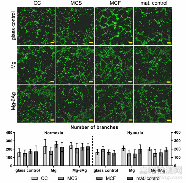
血管生成与内皮细胞增殖和迁移密切相关。因此,可降解镁基材料条件培养基共培养可监测HUVEC的增殖和迁移。图8a总结了直接改变条件培养基第0天、3天和7天后,由细胞核染色归一化荧光强度显示的HUVEC增殖情况。在所有条件下,对照组中均观察到稳定的HUVEC增殖,HUVEC与含Mg (CC, MCS, MCF)培养基共培养基孵育后,内皮细胞增殖从第0天到第3天显著增加,随后在第7天下降,而在Mg-6Ag培养基中只观察到少量细胞数量变化。此外,细胞划痕实验表明含Mg的材料还能明显减少HUVEC的细胞迁移,如图8b所示。
Fig. 8a. Endothelial cell proliferation with different conditioned media. HUVEC were incubated with conditioned media from the coculture (CC) or Saos-eGFP (MCS)and RF Fibroblasts (MCF) in monocultures on Mg-based materials or material without cells. Fluorescence intensities (normalized to day 0) are shown as the mean ±SD from two experiments with six triplicates (n = 12). Statistics: two-way ANOVA (materials, time points; compared to day 0) with Tukey’s multiple comparison test.* = p < 0.05; ** = p < 0.01; *** = p < 0.001.
Fig. 8b. Endothelial cell tube formation. Representative pictures of HUVEC capillary structures under normoxia stained with calcein-AM. Scale bar is 100 μm. Imageswere analyzed with the “Angiogenesis analyzer” from ImageJ and branch numbers are shown as the mean ± SD from two experiments with three samples (n = 6).Statistics: two-way ANOVA (materials, time points) with Tukey’s multiple comparison test.
图9证明了在常氧(图9B)和缺氧(图9C)条件下,在不同条件介质中孵育48 h后HUVEC的迁移(图9A)。常氧条件下,含镁材料并不影响HUVEC的迁移,但翻样条件下,可显著降低HUVEC的迁移,从而抑制肿瘤相关血管生成。
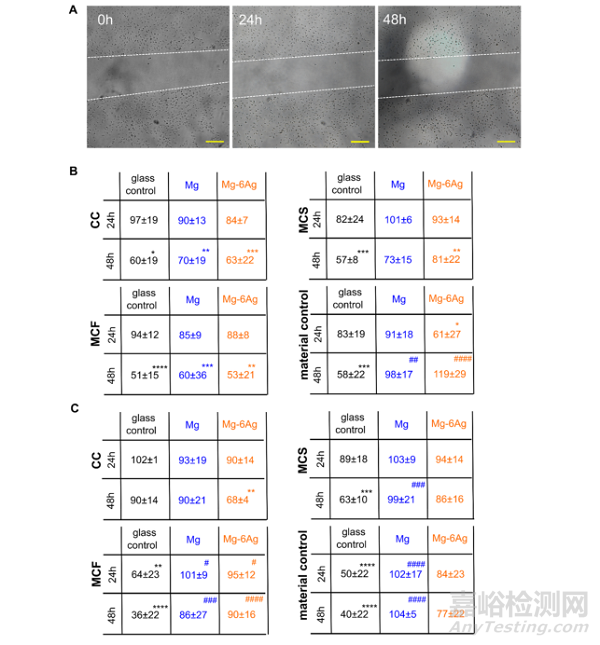
Fig. 9. Endothelial cell migration with different conditioned media. HUVEC were incubated with conditioned media from the coculture (CC) or Saos-eGFP (MCS) andRF Fibroblasts (MCF) in monocultures on Mg-based materials or material without cells. (A) Representative microscopic images of the scratch area in a HUVEC layerwithin 48 h. The white dotted lines symbolize the cell fronts. Scale bar is 100 μm. (B, C) Quantification of the scratch area in relation to the initial cell-free area undernormoxia (B) and hypoxia (C). Relative cell-free areas are shown as the mean ± SD from two experiments with two samples and three chosen positions (n = 12).Statistics: two-way ANOVA (Mg, Mg–6Ag compared to glass control = #; 24 h, 48 h compared to 100% at 0 h = *) with Tukey’s multiple comparison test. Onesymbol = p < 0.05; two symbols = p < 0.01; three symbols p < 0.001; four symbols = p < 0.0001.
5. 结论
研究结果表明,含镁材料是一种具有抗癌活性的可降解材料,在骨肉瘤的发生和发展中,该材料通过抑制癌细胞的迁移、侵袭和肿瘤相关血管的生成来减少骨肉瘤转移的发生率。然而,含镁基材料降解的决定环境因素包括pH和渗透压等。我们的研究仅揭示了缓慢降解的镁材料对骨肉瘤进展的影响,而未来的研究应使用不同降解率的材料来验证这些结果。
原文信息:Mg-based materials diminish tumor spreading and cancer metastases.
Philipp Globig, Roshani Madurawala, Regine Willumeit-Römer, Fernanda Martini, Elisa Mazzoni, Bérengère J.C. Luthringer-Feyerabend.
Bioactive Materials, 19 (2023) 594-610.