角膜损伤是导致角膜瘢痕形成,影响角膜透明性和功能的常见原因,目前临床需通过角膜移植进行角膜重建。中山大学中山眼科中心袁进教授和材料科学与工程学院白莹副教授在科爱出版创办的期刊 Bioactive Materials 上联合发表研究文章:通过构建互不竞争双交联天然聚合物互穿网络,研发透明度高、良好组织粘附性和细胞相容性的复合水凝胶,促进角膜上皮和基质再生,抑制瘢痕形成,重建透明角膜。
1、研究内容简介
角膜移植虽然是角膜损伤功能重建的有效方法,但是供体紧缺仍然是制约角膜复明手术开展的瓶颈,且供体角膜植片需要通过缝合固定,不仅会带来散光,而且存在如新生血管、缝线感染等诸多并发症。针对局灶性的角膜损伤采用可原位形成组织粘附性和促再生能力的透明水凝胶进行修复成为新的研究方向。
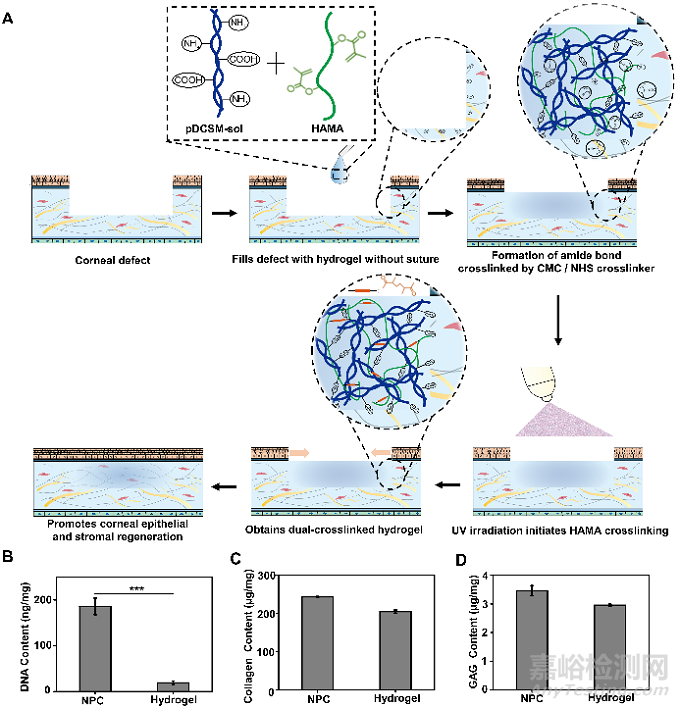
猪来源的去细胞角膜基质水凝胶(pDCSM-G)来源广、可注射。它保留角膜组织中的活性成分,相比单一组分的水凝胶,能够更好模拟原生组织微环境。但pDCSM-G的力学强度较弱、降解很快,无法作为长期有效的角膜修复材料。为了提高pDCSM-G的力学强度和抗降解性能,课题组选用CMC/NHS作为pDCSM-G的交联剂,并通过引入可光交联的甲基丙烯酰化透明质酸(HAMA)得到双交联、双网络复合水凝胶,实现无缝合地快速密封不同形状和大小的角膜缺损,并且可以促进角膜再上皮化及基质再生。
Fig. 1. Application, synthesis, and biomolecular compositions of hydrogels. (A) Schematic illustrations of the hybrid hydrogel applied onto corneal defect in situ for long-term regenerative repair. Quantitative analysis of the (B) DNA, (C) collagen, and (D) GAG contents in the NPCs and those retained in the pDCSM-G, respectively. (***, P<0.001).
双交联复合水凝胶具有理想的光学特性及结构稳定性,其透光率在可见光范围内超过80%。优选成分比例(C3H1)的复合水凝胶在PBS溶液中几乎不发生溶胀,并且光交联HAMA组分的引入赋予该水凝胶较好的抗酶解特性。
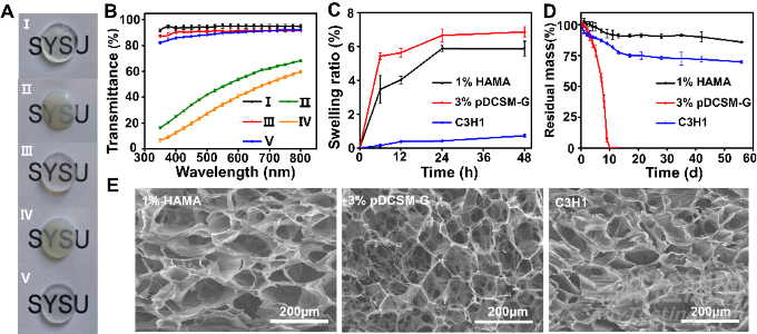
Fig. 2. Optical, swelling, degradation, and microscopic characterizations of the hydrogels. (A) Photographs of the prepared hydrogel specimens (thickness ~ 1 mm) placed on an "SYSU"-labelled paper. The five hydrogel specimens were, (I) photocrosslinked 1% HAMA, (II) physically crosslinked 3% pDCSM-G without CMC/NHS at 37°C, (III) CMC/NHS crosslinked 3% pDCSM-G, (IV) photocrosslinked C3H1 hybrid hydrogel without CMC/NHS at 37°C, and (V) dual-crosslinked C3H1 hydrogel. (B) Light transmittance of the abovementioned five hydrogel specimens on visible light spectrum. (C) Swelling ratios of the photocrosslinked 1% HAMA, CMC/NHS crosslinked 3% pDCSM-G, and dual-crosslinked C3H1 hydrogels when immersed in PBS solution at 37°C in PBS solution within two days, respectively. (D) Long-term degradation behavior of the photocrosslinked 1% HAMA, CMC/NHS crosslinked 3% pDCSM-G, and dual-crosslinked C3H1 hydrogels in 5U/mL collagenase type I solution at 37°C within 60 days, respectively. (E) SEM micrographs of the lyophilized photocrosslinked 1% HAMA, CMC/NHS crosslinked 3% pDCSM-G, and dual-crosslinked C3H1 hydrogels.
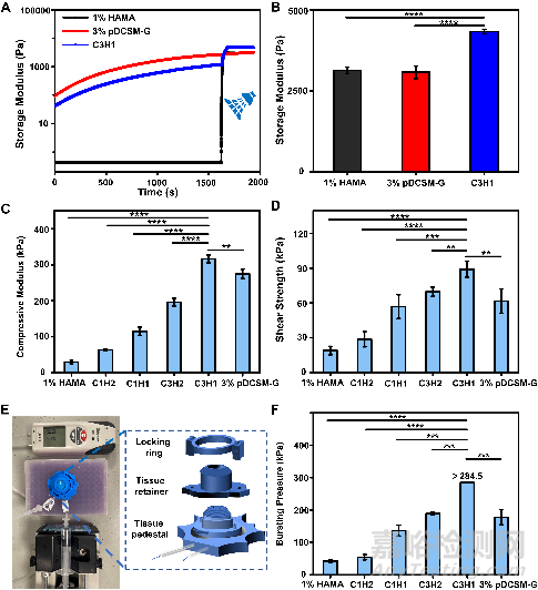
在水凝胶力学性能测试中,流变测试显示该复合水凝胶结合了pDCSM-G和HAMA水凝胶的成胶特性。优选成分比例(C3H1)的复合水凝胶拥有最佳的抗形变性能和组织粘附性,在离体猪角膜的压力测试中,它可承受超过284.5mmHg的眼内压。这些性质将有利于复合水凝胶在无缝合情况下长期稳定地黏附在基质植床上。
Fig. 3. Mechanical characterizations of the chemically crosslinked hydrogels. (A) Rheological characterization of the photocrosslinked 1% HAMA, CMC/NHS crosslinked 3% pDCSM-G, and dual-crosslinked C3H1 hydrogel before and after light exposure. (B) storage moduli of the 1% HAMA, 3% pDCSM-G, and C3H1 hydrogels after chemical crosslinking, characterized by rheometry. (C) Compressive moduli of the hydrogels with different compositions of HAMA and pDCSM-G. (D) Shear strength of the hydrogels with different compositions of HAMA and pDCSM-G. (E) Photograph of the artificial anterior chamber device used for bursting pressure tests. (F) Averaged bursting pressures of the hydrogels with different compositions of HAMA and pDCSM-G. (**P<0.01, ***P<0.001, ****P<0.0001)
体外细胞培养证明:复合水凝胶无细胞毒性,有利于角膜细胞的黏附、存活和增殖,且能够帮助维持角膜基质细胞的稳定型,减少角膜基质瘢痕形成。
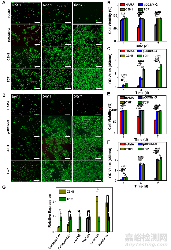
Fig. 4. Corneal epithelial cells and corneal stromal cells cultured on the HAMA, pDCSM-G, C3H1 hydrogels, and TCP. (A) Live/Dead staining of corneal epithelial cells cultured on 1% HAMA, 3% pDCSM-G, C3H1 hydrogel, and TCP for 1, 4, and 7 days, respectively. Scale bars = 200 μm. (B) Quantification of the corneal epithelial cells viability on hydrogels and TCP characterized by Live/Dead assay. (C) CCK-8 assay of the cultured corneal epithelial cells after 1, 4, and 7 days. (D) Live/Dead staining of corneal stromal cells cultured on 1% HAMA, 3% pDCSM-G, C3H1 hydrogel, and TCP for 1, 4, and 7 days. Scale bars = 200 μm. (E) Quantification of the corneal stromal cells viability on hydrogels and TCP characterized by Live/Dead assay. (F) CCK-8 assay of the cultured corneal stroma cells after 1, 4, and 7 days. (G) qRT-PCR analysis of the relative mRNA expression levels of Collagen 1A1, Collagen 3A1, ACTA2, TGF-β1, lumican, and keratocan in the rabbit corneal stromal cells after culturing on the C3H1 hydrogel and TCP for four days. (*P<0.05, **P<0.01, ***P<0.001, ****P<0.0001)
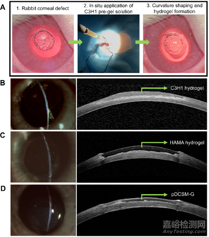
在新西兰大白兔基质缺损模型中,该复合水凝胶能够简单快速地被注射到基质缺损处,通过自发的CMC/NHS交联剂交联和HAMA光引发成胶,可以无缝合、无缝隙地停留于缺损处,展示出操作的实用性和便捷性。裂隙灯和AS-OCT结果显示,刚填充后的水凝胶能够与基质植床紧密贴合,角膜厚度和曲率得到一定的恢复。
Fig. 5. In vivo application of the hydrogels onto rabbit corneal defects. (A) Procedure of hydrogel injection onto the corneal defect. Representative slit lamp photographs (left) and AS-OCT images (right) after hydrogel treatments using (B) C3H1 hydrogel, (C) 1% HAMA hydrogel, and (D) 3% pDCSM-G, respectively.
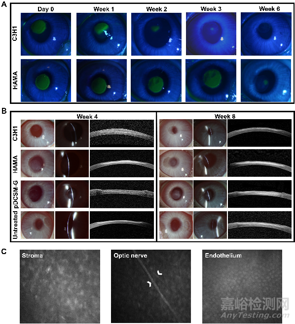
原位植入八周后,填充复合水凝胶的实验眼在修复前期能够实现缺损区域的再上皮化,角膜保持良好的透明度和角膜弧度,没有明显的炎症反应和瘢痕生成。复合水凝胶的填充不会对角膜神经、基质层或内皮层产生不利影响。其修复效果明显地好于无法实现快速再上皮化的HAMA组和过快降解的pDCSM-G组,这两组的实验眼均有明显的基质瘢痕形成。
Fig. 6. Postoperative observation of the hydrogel treated corneas. (A) Representative photographs of cobalt blue with fluorescein staining in the rabbit experimental eyes filled with C3H1 and 1% HAMA hydrogels at different time points. The green area in the central cornea indicates the epithelial defect. (B) Representative slit lamp and AS-OCT images of the experimental eyes filled with C3H1 hydrogel, 1% HAMA hydrogel, 3% pDCSM-G, and the untreated eyes 4 and 8 weeks after surgery. (C) Confocal micrographs of the corneal stroma, optic nerve, and endothelium in the C3H1-hydrogel-filled cornea 8 weeks post-operation. White arrows point at the optic nerves of the posterior stroma.
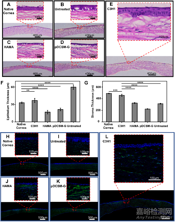
在所有实验组中,复合水凝胶在填充至角膜缺损处八周后,得到最接近正常健康角膜(native cornea)的上皮层和基质层厚度,基质排列有序,厚度得到恢复。与之相比,HAMA组和pDCSM-G组的缺损处均只生成了单层的上皮细胞层,有明显的基质缺损存在。无处理组则伴随有明显的上皮肥大现象,基质缺损明显。免疫荧光染色结果表明,复合水凝胶组的α-SMA的表达显著低于HAMA组和pDCSM-G组,意味着更少的基质瘢痕。因此,复合水凝胶经缺损处无缝合填充后,表现出更为理想的功能性修复效果和促进角膜再生能力。
Fig. 7. Histological analysis of the hydrogel treated rabbit corneas eight weeks post-operation. Representative H&E images of (A) native corneas without defect, (B) corneas with untreated defect, and the defected corneas filled with (C) 1% HAMA hydrogel, (D) 3% pDCSM-G, and (E) C3H1 hydrogel. Quantification of (F) epithelium thickness and (G) stroma thickness according to the results from H&E staining. Representative images of immunofluorescence stained (H) native corneas without defect, (I) corneas with untreated defect, and the defected corneas filled with (J) 1% HAMA hydrogel, (K) 3% pDCSM-G, and (L) C3H1 hydrogel, using biomarkers α-SMA (green) and DAPI (blue). (*P<0.05, ****P<0.0001)
综上所述,该研究利用互不竞争的双交联体系构建的复合水凝胶展现出良好的理化性能和促再生生物活性,高透明度、良好稳定性、可控机械性能及黏附性能,支持角膜细胞的生长和维持修复表型。该新型材料可简便快捷地填充不规则的角膜缺损,实现角膜快速再上皮化,促进角膜基质再生、减少角膜瘢痕,表现出良好的功能性修复效果和促进角膜再生能力,为角膜创伤修复的治疗提供了新的干预模式。