您当前的位置:检测资讯 > 科研开发
嘉峪检测网 2022-03-03 06:06
组织工程支架提供有利于细胞生长的微环境,是组织再生中的重要组成部分。各向异性支架模拟了神经和脊髓的细胞外基质结构,可以有效引导细胞轴突和雪旺细胞定向生长。该综述首先介绍了外周神经和脊髓的解剖结构以及损伤的临床和潜在治疗方法,并从一维的表面特征、二维的纤维基材和三维的水凝胶三个方面综述了目前各向异性神经/脊髓再生支架的制备和研究进展。
01、研究内容简介
脊髓损伤(SCI)伴随着一系列的细胞反应,在损伤发生部位,神经元和胶质细胞死亡,血管发生破裂(Figure 1A)。之后炎症细胞聚集,胶质疤痕形成以保护损伤部位。随着炎症反应降低,脊髓通过轴突再生和新血管形成开始自我修复。但胶质疤痕的细胞外基质,包括硫酸软骨素和蛋白聚糖阻止了神经元的有效再生。
外周神经损伤(PNI)后,远端的轴突脱髓鞘化并且发生降解,这一过程也称作Wallerian degeneration (Figure 1B)。之后近段形成Büngner bands促进神经再生,包括长度方向取向的雪旺细胞引导轴突向远端生长,过程中也伴随着炎症反应。对于神经元没有完全断裂的损伤,神经可以通过自我修复完成再生。但对于大段神经损伤,炎症细胞、基质的纤维化等使得神经损伤无法恢复。
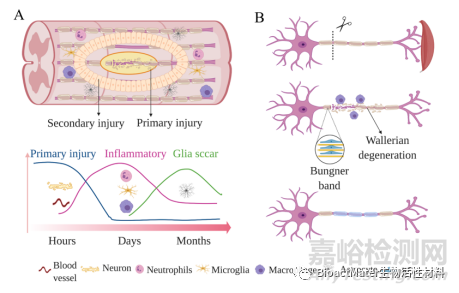
Figure 1. Physiological changes after SCI (A) and PNI (B) at the injury site
各向异性的一维表面包括连续性和非连续性表面特征,其通过“接触导向”作用影响着附着细胞的行为。制备通常以不可降解的硅、聚苯乙烯、PDMS和可降解的PLA,PCL等为基材,制备方法主要为激光刻蚀、光刻和软光刻 (Figure 2)。凹槽和脊状是典型的连续性表面特征,其宽度和深度影响着附着细胞的取向度。有研究表明,当凹槽的宽度和深度接近细胞尺寸时,雪旺细胞能更好的沿着凹槽方向生长,且上表达有关细胞骨架、髓鞘化等基因。作为非连续性的分布式柱状和点状特征,其各向异性排列,以及表面粗糙度也影响着DRG 和交感神经元的轴突生长。
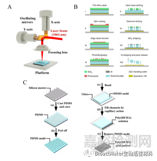
Figure 2. A) Schematic of laser scanning. B) Schematic of photolithography. C) Four major soft lithographic techniques.
静电纺丝纤维基支架较大的比表面积和孔隙率,使其广泛应用于组织工程支架。作者总结了用于制备各向异性外周神经和脊髓再生支架的静电纺丝接收装置,包括旋转的盘状、柱状、轮状、杆状以及平行电极等。为了弥补静电纺丝支架力学性能等不足,一些纤维自组装、剪切力/拉伸力、相分离等技术也被用于制备各向异性的外周神经和脊髓再生支架,作者在文中对其制备原理进行了综述。各向异性纤维基材料可成膜状,并进一步卷成管状,或直接制备成管状,以及制备成纤维束作为填充物,来连接损伤神经。雪旺细胞和轴突能沿着纤维方向生长和迁移,有效促进神经的再连接(Figure 3)。通过结合不同尺度的各向异性,如纳米级和微米级,神经细胞的再生可以进一步被提高。
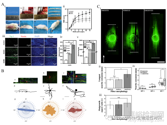
Figure 3. A) i: Pictures of animal experiments. a: Image of the electrospun GelMA hydrogel fiber scaffold. b: 3 mm hemisection made in the right site of the T9 spinal cord. c: Scaffold implantation. d: Spinal cord specimens were collected 12 weeks after surgery. From top to bottom: control group, gelatin group, GelMA group. e–h: Animals at weeks 1,4,8, and12 after surgery. ii: Evaluation of the lower limb motor function of rats with a BBB score. iii–v: Immunofluorescence staining of neural stem cells and neural cells and the quantitative comparison of optical density among groups. Samples without implants were used as a control group. *p < 0.05. B) a–c: 40× fluorescent images of individual neurons seeded on astrocytes, which were cultured on aligned fiber scaffolds (a), PLLA films (b), or fiber/AFFT scaffolds (c). d–f: Isolated traces of individual neurons pictured in a, b, and c, respectively. g–i: Polar histograms showing total outgrowth and orientation of neurites seeded on astrocyte layers, which were cultured on the three scaffold types (g = aligned fibers, h = film, i = AFFT boundary). C) DRG extension (green: ß tubulin) on SAS fibers exhibiting smooth, porous, and grooved topography (i). Grooved fibers demonstrate a significantly longer neurite extension compared to smooth and porous fibers after DIV 7 (ii). Effect of fiber surface topography on DRG surface area aspect ratio after DIV 7 (iii). Neurite extension length from DRGs on fibers after DIV 1, DIV 4, and DIV 7 (iv). Scale bar is 1 mm. The white arrow shows fiber direction.
用于外周神经/脊髓的各向异性水凝胶具有巨大的应用潜力,作者总结了常用的制备方法,详述了制备原理,其中包括单向冷冻法、3D打印、离子扩散法、磁场/电场,外力作用,以及模具法(Figure 4)。通过控制水凝胶支架的孔径,可以得到曲向的微观或宏观的孔和通道。同样的通过“接触导向”作用,曲向的水凝胶孔或通道可以促进胶质细胞和轴突的定向生长,进而加速神经再生过程。
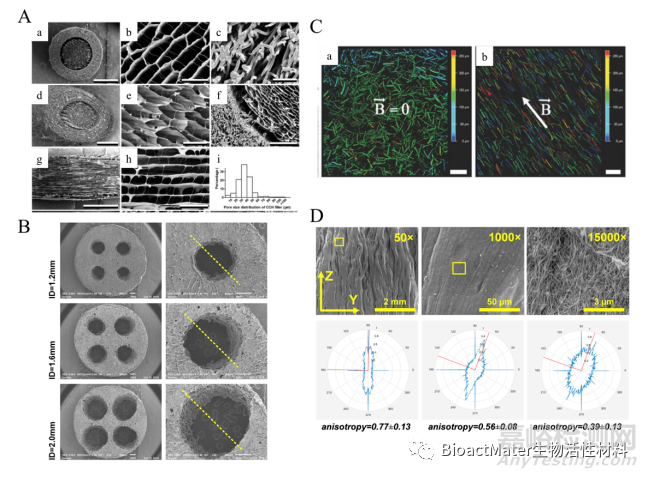
Figure 4. A) SEM characterization of the scaffold. a-f: Transverse section of the scaffold (a, d) with collagen/ chitosan filler (b, e) and PCL sheath (c, f). G–H: Longitudinal section of the collagen/ chitosan filler. i: Pore size distribution of the collagen/ chitosan filler. a, d, g: scale bars = 1 mm; b, c and e: scale bars = 50 μm; f: scale bar = 200 μm; h: scale bar = 100 μm. B) Scanning electron microscopy images of nerve guidance conduits showing the transverse section (dotted yellow line of the image on the right showing the plane where the samples were cut to acquire longitudinal images. C) Depth color‐coded images of magnetic fibers inside 3D fibrin hydrogels, prepared a: in the absence of an external magnetic field and b: in the presence of a 100 mT magnetic field. D) Multiscale anisotropy of biaxially compressed collagen scaffolds. The top row shows SEM images at the magnification specified in top right corner. The bottom row shows the corresponding polar plot from the image analysis, where the red lines represent the primary fiber axis, and the blue line is the magnitude of alignment at the specified angle.
最后,作者从生长锥和粘着斑两个角度介绍了细胞和轴突在各向异性支架中定向生长的机理。生长锥是生长的轴突前端的感应性结构,由肌动蛋白纤维和微管组成(Figure 5A),它们控制着生长锥的前进、后退等行为。束状的微管在生长锥的中心部位,外周为肌动蛋白构成的板状伪足和丝状伪足。板状伪足和丝状伪足能感应、识别外界形貌和压力的变化,并使自身沿着各向异性形貌曲向,以减少细胞骨架变形带来的压力。微管结构能剧烈的收缩和运动,进一步控制细胞的运动。粘着斑由大量的整合素组成(Figure 5B),整合素是一种跨膜蛋白,连接着细胞骨架和基质,细胞可以通过整合素来根据外界机智改变细胞形态。

Figure 5. Schematic of cellular response to ECM by the growth cone (A) and focal adhesion (B)

来源:Internet